

Diabetes and the eyes: when and how often to see an eye doctor, and what you need to notice yourself
For well-controlled diabetics, the eye often does not come first among the risks. HbA1c, blood pressure, cholesterol, the feet, the kidneys: these topics dominate the diabetic consultations. The eyes come somewhere after that, with a casual „that too,“ often mentioned only in passing.
The most common eye complication of diabetes, diabetic retinopathy, is one of the leading causes of preventable blindness in working-age adults worldwide. And it is precisely because of this that it is so dangerous, as it causes no symptoms in most patients for a long time.
The proportion of treatable eye complications would be surprisingly high if all diabetics were examined in a timely manner. However, the literature shows that 40 to 60 percent of diabetes patients do not receive the recommended eye examinations with the intended frequency. The chain of general practitioner, diabetologist, and ophthalmologist does not close tightly enough. This article discusses what actually happens in the eye with diabetes, which symptoms are warning signs, how often an eye examination is sensible, and what to expect during such an examination.
In this article
Why the eye in particular?
Diabetes is primarily a disease of the small vessels. A persistently elevated blood sugar level damages the capillaries, the smallest blood vessels in the body, in the long term. In the kidney, this leads to kidney failure. In the peripheral nerves, it leads to neuropathy. In the eye, it leads to a very characteristic cascade of changes, which is collectively referred to as diabetic eye disease .
At the back of the eye wall, on the retina, these capillaries supply the light-sensitive sensory cells. Under the influence of high sugar levels, the vessel walls thicken, they expand, and at certain points, they become permeable. This is the first stage, the so-called non-proliferative diabetic retinopathy (NPDR). At various points on the retina, small, dot-like hemorrhages become visible, as well as yellowish lipid deposits (exudates) and milky white, fluffy spots (cotton-wool spots). The ophthalmologist recognizes this immediately with the slit lamp or on the retinal image. The patient usually does not notice anything at this stage.
If the process continues, entire areas of the retina remain without adequate blood and oxygen supply. The body tries to help and allows new, but fragile vessels to grow to close the supply gaps. This is the proliferative diabetic retinopathy (PDR). These new vessels are structurally unstable, bleed easily, and the accompanying connective tissue can pull the retina away from the back of the eye. From here, vision can deteriorate dramatically from one day to the next.
In the middle of the course, often regardless of whether the retinopathy is in the NPDR or PDR stage, another complication can arise: the diabetic macular edema (DME). The macula is the center of the retina and is responsible for sharp, central vision. If fluid leaks into the tissue here and the macula swells, central vision becomes blurry. Statistically, DME is the most common cause of diabetic vision deterioration.

Diabetes affects not only the retina
Diabetic retinopathy is the most significant, but not the only ocular complication. In diabetics, the cataract develops on average three to five years earlier, and often with a faster progression. The elevated blood sugar level chemically reacts with the lens proteins, causing the lens substance to become cloudy over time.
The risk for glaucoma is also increased. In the diabetic population, the likelihood of elevated intraocular pressure is about twice as high as in the non-diabetic population. A particularly aggressive form, neovascular glaucoma, can develop as a complication of severe, untreated diabetic retinopathy when the fragile new vessels also invade the anterior segment of the eye.
Dry eyes are also more common. Diabetic neuropathy can affect the nerve endings of the cornea, reduce the blink reflex, and decrease the sensitivity of the eye surface. For many patients, this manifests as a small but persistent complaint.
And finally, what makes diabetes patients particularly sensitive during any ophthalmic procedure: healing is slower. Corneal wounds scar slowly, and the risk of infection is increased. This applies equally to cataract surgery, laser procedures, and eyelid surgeries.
The symptoms you need to perceive yourself
The most important and at the same time unpleasant message: the early stages of diabetic retinopathy are almost completely asymptomatic. As long as the macula is not affected, there is no noticeable vision deterioration. Small peripheral bleedings at the edge of the visual field go unnoticed in everyday life. The patient receives no warning that a process is already underway in the eye. This precisely justifies the need for regular check-ups, even without symptoms.
When symptoms occur, they are typically the following.
Blurred, fluctuating vision. An elevated blood sugar level changes the refractive power of the eye lens, and with poorly controlled diabetes, vision can fluctuate from day to day. This can be an early sign that blood sugar control is not stable, even before the retina suffers permanent damage.
Blurriness in the central visual field, especially when reading. This is the typical symptom of a diabetic macular edema. Not sharply blurred, but the text „melts“ slightly, the letters appear unclear, reading requires more light.
Black dots, spots, or streaks in the visual field. In diabetes, floaters often do not mean the harmless posterior vitreous detachment, but rather a small actual bleeding in the vitreous body. If new floaters occur in a diabetic, a timely ophthalmological examination is indicated.
A dark or grayish spot in the visual field, often in one sector. This already indicates a larger bleeding or a local damage to the retina.
A sudden, significant deterioration in vision in one or both eyes. The most severe symptom that requires immediate, possibly retinal surgical intervention. Causes can be a vitreous hemorrhage, a retinal detachment, or an acute neovascular glaucoma crisis.
Light flashes and distorted lines, especially when reading. The distortion of straight lines (metamorphopsia) indicates swelling of the macula. If a horizontal line appears curved with one eye and straight with the other, a prompt examination is necessary.
How often for eye examinations?
The international recommendations are based on the type of diabetes, the duration of the disease, and individual risk factors. The guidelines of the American Diabetes Association (ADA), the American Academy of Ophthalmology (AAO), and the Swiss Society for Endocrinology and Diabetology (SGED) largely agree.
Recommended control intervals
Type 1
First examination within 5 years
After diagnosis. Then annual check-up.
Type 2
Immediately upon diagnosis
Because the disease often exists unnoticed for years. Then annually.
Pregnancy
Before conception and in the 1st trimester
Further rhythm according to ophthalmological guidelines.
In case of existing retinopathy or unstable blood sugar levels: closer controls according to individual medical recommendation.
More frequent checks are necessary if retinopathy has already been diagnosed (depending on severity every three to six months), in case of unstable blood sugar levels, with rapidly changing HbA1c values (e.g., after newly initiated insulin therapy), in renal insufficiency, or severe hypertension.
In general practice and diabetology, according to international data, the most common gap is here: the patient has been treated for years, HbA1c values are regularly measured, but an ophthalmological check has not taken place in the last three to four years. This is not an exception but the typical form of the problem.
What to expect during a diabetic eye examination
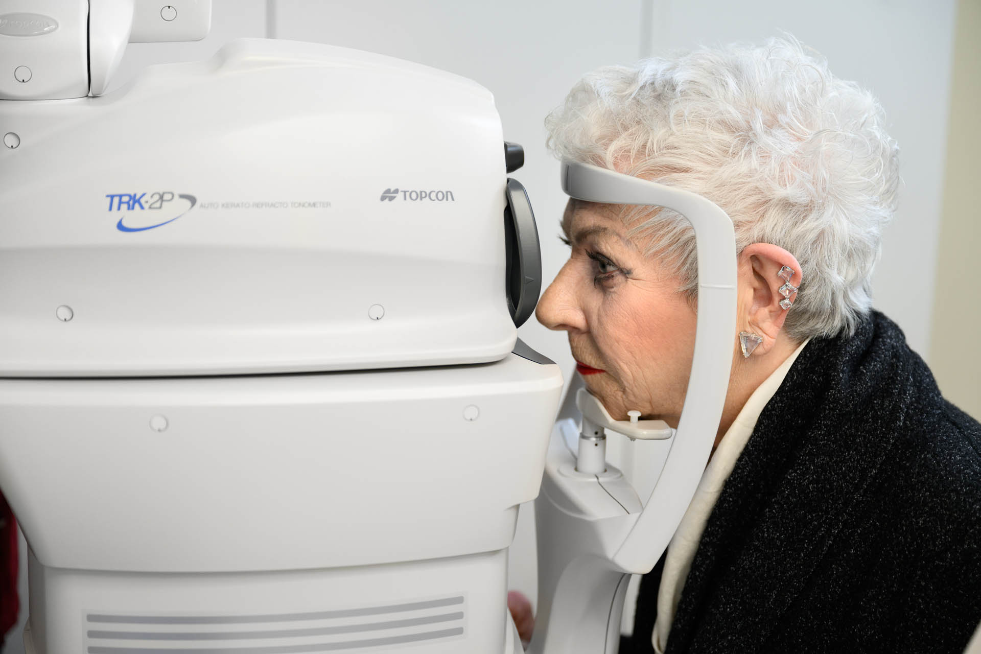
The diabetic ophthalmological examination is essentially an extended retinal examination, supplemented by imaging techniques that document the condition of the macula and retinal layers.
The first step is the measurement of visual acuity and intraocular pressure. It is followed by the pupil dilation: with eye drops, the pupil is dilated within twenty to thirty minutes, allowing detailed examination of the peripheral retinal areas. The peripheral signs of retinopathy appear first in these marginal areas, which is why pupil dilation is not optional.
The fundus examination is performed with an ophthalmoscope or with a slit lamp. The diabetic changes such as microaneurysms, small hemorrhages, exudates, cotton wool spots, and new vessels show a typical pattern.
In modern practice, two imaging techniques have become standard. The optical coherence tomography (OCT) shows the retinal layers in cross-section, with micrometer precision. The macular edema can be clearly documented with the OCT. The fundus photography or wide-angle retinal imaging captures the entire retina in a single image and allows for precise longitudinal comparison.
In case of suspicion of proliferative retinopathy or diabetic macular edema, supplementary procedures such as fluorescein angiography (FAG) or OCT angiography (OCTA) are useful.
After the examination, near vision is blurred for a few hours, and light sensitivity is temporarily increased. It is advisable not to drive back yourself and to bring sunglasses.
Adlerblick Eye Medicine · Döttingen
The diabetic eye examination
Once a year, 45 minutes, a clear report. The easiest way to protect vision in the long term.
Treatment options
The treatment of diabetic eye complications has changed revolutionarily in the last fifteen years. What could previously only be slowed down with lasers can today be reversed in many cases with modern medications, sometimes completely treated.
The Anti-VEGF injections (Aflibercept, Ranibizumab, Bevacizumab, Faricimab) are the first-line therapy for diabetic macular edema. The injection is administered directly into the vitreous body, on an outpatient basis in the practice. The medication reduces vascular permeability, the macular fluid is absorbed, and vision improves. Treatment is done in series, usually six to eight injections in the first year.
The Retinal laser therapy, the pan-retinal photocoagulation (PRP), is the standard therapy for proliferative retinopathy. The doctor places controlled laser spots in the peripheral retinal areas that deactivate the hypoxic areas and stop neovascularization. The procedure is outpatient and typically spreads over two to three sessions.
In severe cases with pronounced vitreous hemorrhage or traction-related retinal detachment, the vitrectomy (pars plana vitrectomy) is considered as a surgical intervention. This procedure is performed in retinal surgical centers.
For all forms of treatment, the earlier the complication is detected, the better the outcome. This is the only reason why prevention is so crucial.
What you can do yourself
In addition to medical treatment, personal lifestyle factors are at least equally important.
What you can influence yourself
- Blood sugar control: a reduction in HbA1c by 1 to 2 percentage points lowers the risk of retinopathy in the long term by 20 to 40 percent.
- Blood pressure control: Target value usually below 130/80 mmHg, depending on individual risk profile.
- Cholesterol and triglyceride control: reduces lipid deposits (exudates) on the retina.
- Smoking cessation: Smoking significantly accelerates the vascular complications of diabetes.
- Exercise and balanced diet: Components of basic diabetic treatment, with direct benefits for the eyes.
Regular eye examinations do not directly prevent retinopathy, but they ensure that changes can be detected and treated in a timely manner. Prevention and screening are two different things; both are important.
Frequently asked questions
Question
If my blood sugar is well controlled, do I still need to see an eye doctor?
Yes. Good blood sugar control significantly reduces the risk of retinopathy, but it does not eliminate it. Annual eye examinations are also indicated when the HbA1c level is within the target range. Certain forms, such as macular edema, can occur even with well-controlled diabetes.
Question
When should I see an eye doctor for the first time after a new diagnosis?
For type 2 diabetes, immediately after diagnosis. For type 1 diabetes, within five years after diagnosis. For diabetes that occurs very early in childhood, the first examination is recommended at the onset of puberty.
Question
If I have diabetes and suddenly experience floaters, is that urgent?
Yes. In diabetes, new floaters, streaks, or even dark shadows in the field of vision are not harmless phenomena. They often indicate a small vitreous hemorrhage or proliferative retinopathy. An eye examination within a few days is indicated.
Question
Can my vision worsen during pregnancy?
Yes. The hormonal and circulatory changes of pregnancy can accelerate diabetic retinopathy. If you are planning to have children and in the first trimester, an eye examination is advisable; further appointments will be scheduled by the specialist.
Question
Does an anti-VEGF injection hurt?
It is not painful. The eye doctor numbs the eye with drops, and the injection is given through the white, less sensitive part of the eye. The injection takes only a few seconds. Afterwards, there may be a slight foreign body sensation for a few hours, which quickly passes.
Question
What to do in case of sudden vision deterioration?
An eye examination on the same day, if possible. An acute, sudden vision deterioration can indicate a vitreous hemorrhage or retinal detachment in diabetes. Immediate clarification significantly affects the final outcome.
What matters
Diabetes is a systemic disease that affects the entire body, and the eyes are no exception. The good news is that the vast majority of diabetic eye complications can either be prevented or effectively treated if detected in time. The difficult message is that the early stages are asymptomatic and do not come to light without active screening.
„Vision does not return once it is lost. If the risk is contained in time, most diabetes patients can retain what they have in front of their eyes today.“
If you live with diabetes and have not had an eye examination in the last twelve months, now is the time. If you notice new symptoms, such as blurred or fluctuating vision, floaters, unclear central areas, or a sudden deterioration in vision, do not wait until your next diabetes appointment, but make an appointment with the eye doctor.
Adlerblick Eye Medicine in Döttingen, in the center of the canton of Aargau, offers complete diabetic eye diagnostics: extended retinal examination, OCT, fundus photography, and, if necessary, referral to the appropriate retinal surgical center for the treatment phase. Our focus is on prevention and early detection, as this offers the best chance to preserve vision.
The examination is simple, painless, and the effort of once a year is nothing compared to what is at stake.